Brain Imaging, Crash Course ️

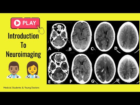
hello and welcome to our last presentation today we talking about neuroimaging okay fast reviews of our goals and objectives for this lecture you always have to know the anatomy very well you know anatomy you cannot learn radiology uh you have to know what this mri is more appropriate or for spine imaging or why the CT scan is more appropriate for emergencies you have to discriminate between different pathologies for the brain, we're talking about emergencies mainly and you have to construct the appropriate imaging approach of common diagnosis okay for start let's go to talk about what is a neuroimaging an umbrella term encompassing a variety of methods and technologies okay it was divided into main areas: structural imaging and functional imaging so today we have we focus about extruder imaging okay that the tutorial imaging provides image of the brain anatomy structure. this type of imaging helps in the diagnosis of brain injury and the diagnosis of certain diseases okay in this case we use a CT scan and MRI remember just we want to look at the functional imaging Functional imaging provides images of the brain image as patients complete tasks such as solving math problems or reading or responding to stimuli such as auditory sounds or flashing lights. examples of these ones are positron emission tomography regional cerebral blood flow, and functional magnetic resonance imaging this is a sample of functional structure so in this case this is in 3D with a lot of colors there we can find thIS kind of studies in big institutions and research institutions in a few countries so let's be focused on our issue today, structural imaging so let's go to talk about computer tomography and MRI, so uh Computer Tomography uses a lot of radiation okay remember, MRI doesn't use radiation, for that reason, it's very safe for obstetric patient but what happened is, the computer tomography can be very fast, you can scan a patient I think the head of the patient is less than one minute for any type of study of MRI should be between 20 to 25 minutes MRI is very very good for soft tissues. It going to give you more details for soft tissues and the spine CT Scan because it's faster is very good for emergencies, because it's faster is very good for emergencies, for any head trauma, we don't request MRI, we request a ct scan okay because it's very fast, you cannot put a patient with head trauma for 25 -30 minutes inside of the MRI machine, they can die over there but for ct scan remember sometime in less than one minute okay so remember ct scan findings i will described densities and MRI intensities densities and MRI intensities okay now let's go and be focus in emergencies, are the things you are going to be seeing more during your career unless you become radiologists okay so let's go to talk about ct scans. these are relative densities, remember our first lecture, the bones has a high density and the air a low density so the air is hypodense, everything with a gray soft tissue and great it should be isodense and the brightness is high density is hyperdense, now how to describe a head CT scan you have always to start with this sentence. this is a non-contrast or contrast enhanced maybe axial, sagittal, or coronal head CT scan showing. you always have to start with
this one, we have a simple ct scan which is a non-contrast and contrast CT scan we can use both so for a contrast CT can we use the iodine compounds which are positive you remember that so this is an example of normal CT scan from top to bottom of course you have to look at it and you have to go for the anatomy, remember we talked about anatomy in our first lecture, I think, you have to go and look at the anatomy normal axial head CT scan. you have to go and deep on that, also you have to look at the distribution of artery's territory. is a distribution of the anterior cerebral artery middle artery and posterior cerebral artery. you have to look at it very deep now let's go to talk about lesions, for defined lesions you have to know the relationships and location within the brain tissue. intraparenchymal is located within brain tissue. the extraparenchymal is located within the bony casing of the central nervous system but outside the brain tissue itself all right so let's go to find out these locations and we are going to start with extra parenchymal locations, you can see it here we have this two examples of Meningiomas, okay which are benign tumors, these are extra parenchyma location the tumor this kind of tumors is slow growing but you can see is reflecting a mild displacement of the other brain structures, and you see is growing from outside of your brain tissue there are a few characteristics of differences more between the intra and extra parenchymal brain location now this is an example of intraparenchymal lesions we had the example of Neuroblastoma, you see they are in the middle of the brain.
remember you have to look at it, what if the difference between intra and extra parenchymal locations, okay inside of brain pathologies. Now Head CT in acute situations. anytime someone has head trauma, they should have head ct scan or repeat again, anytime someone has head trauma you have to request for ct scan in this case, you are looking for acute blood, which is bright or hyperdense or so all medical doctors should know what acute blood looks like and be able to describe it and it is general location, there are five general locations for acute blood we have Epidural, Subdural, Subarachnoid, Intraventricular, intraparenchymal These are the anatomic specification for what i explained to you and these are examples of bleeding okay in the first one you have an extradural hematoma in the other, we have the middle shift we got intraparenchymal hematoma which is inside of the brain then we have a subdural hematoma which is very very dangerous the patient can die from a subdural hematoma and subarachnoid hemorrhage. you can see the locations. now let's go to be specific about this one subdural hematoma, you see there is a left subdural hematoma which is shifting the brain to the right producing mass effect you can see there the left lateral ventricle is compressed because of this hemorrhage and is shifting the line to the other side they will call it mass effect, mass effect.
you have to know very well the difference between extradural hematoma and subdural hematoma know very well you have to go deep and know the difference between extradural and subdural these are other examples of multiple locations of this kind of hemorrhage, these are contusion hematomas, you can see all these extradural haematomas, some intraparenchymal after trauma The management of the patient depends on the Glasgow of the patient and also depends of the amount of blood, the management of the patient depends on the Neurosurgeon, quantity, position and the Glasgow of the patient so this is another example of being different location you have an intraventricular hemorrhage this is another example of bleeding, we have a subarachnoid hemorrhage remember you have to read everything what i put in the slides on this video to stop the video and read everything if i told you go deep in this area you have to go deep okay you have to review later to be like your homework now how to assess a CT of the head before you call a Neurosurgeon, you have to ask yourself, these six main questions, these are the questions I do myself every day to read a CT scan the First question is if the brain is in the middle of the head? do the 2 sides of your brain look alike? is the fourth ventricle in the midline and symmetrical? Are the lateral ventricles huge with effaced sulci? Are there any abnormal white things inside the skull? or are they any abnormal black things inside the skull? remember white things hyperdense and black things are hypodense, so if there is any abnormal hyperdense thing inside the skull? or there is any abnormal hypodense inside the skull? okay so first question is the brain in the middle of the head? you can compare the left side which is normal with the right side, you can see the brain is not in the middle of the head. do the two sides of the brain look-alike? you can see, in the left side, both sides look-alike but in your right, it doesn't look alike if the fourth ventricle in the midline and symmetrical? you can see in your left , the four ventricle in the middle line and symmetrical and hypodense. in the midline of the posterior fossa, but you can see in the second image, is not in the middle line and there is no hypodense, there is hemorrhage, and then the other is a tumor ,there is a tumor so talk about the ventricles you can see two different 30 years old patient in the left is a normal ventricle size in the right side there is a ventriculomegaly The wideness of the ventricle is up 1,0 centimeter in young patients after 60 years up to 1.5 centimeter it's up to 1.5 centimeters so
anything above or that it considers ventriculomegaly okay you have to pay attention to that let's look at this example this is a 68 yrs old female brought in by ambulance after rapid mental status deterioration we consider the hypertensive hemorrhage, in this case, there is no trauma okay and the head CT scan shows, you can see. the patient has a history of high blood pressure and you can see this intraparenchymal hemorrhage or hematoma huge hematoma you have this hyperdense area surrounded by hypodense area which is edema with the shifting of the middle line to the right side to the right side okay that means there is a huge mass effect this is the final diagnosis. the left side edema and significant midline shift or mass effect we look at this hypodense thing inside of the brain remember, are there any abnormal black things inside of the brain? when we talk about hypodense things inside of the brain, we talk about stroke we have to think first about the stroke, ischemic stroke in this case, stroke can be hemorrhage like we saw before and ischemic, hemorrhage is vessel rupture and ischemic is vessel obstruction. okay an infarction so this patient developed a headache, completely loss of consciousness and vomiting okay you can see there is a large area of hypodense or low density in the right cerebral hemisphere which represents a right middle cerebral artery infarct. so you have to remember to look at the territory of the arteries.
and this is another example of infarction or stroke, ischemin stroke, there's another example here you can see this hypodense area on both sides one in your left which is in the right, but you can see this one in your left in which the hypodense is in the right you can see the hypodense is huge and there is no shift of the midline and also there is the right lateral ventricle is not compressed. you compare with the left side the lateral ventricle is dilated is deleted because we are in the presence of chronic ischemic stroke or cerebral infarct, that for you you have to go and go deep in what is an acute, subacute, and chronic cerebral infarct so these are examples of kind of edema but in this case we're in the presence of tumor you can see in your left this is a non-contrasted ct scan which just appeared this vasogenic edema but after that we saw we think okay this is look like a stroke but this edema look more like a tumor so we put a contrast inside the patient and you can see this hyperdense ring inside of this hypodense area area, we are in the presence of a tumor, so let's move on. these are examples of other tumors, for your general knowledge but for us I think this is all for today remember you have a few homework to do also reporting more videos so you can understand better because this this is trauma an emergency is key for you is the one that you were to watching almost every day so see you
2021-09-23 04:27


